Home
/ Pelvic Anatomy Posterior / Iliac Screw For Reconstructing Posterior Pelvic Ring In Tile Type C1 Pelvic Fractures Sciencedirect / Über 7 millionen englischsprachige bücher.
Pelvic Anatomy Posterior / Iliac Screw For Reconstructing Posterior Pelvic Ring In Tile Type C1 Pelvic Fractures Sciencedirect / Über 7 millionen englischsprachige bücher.
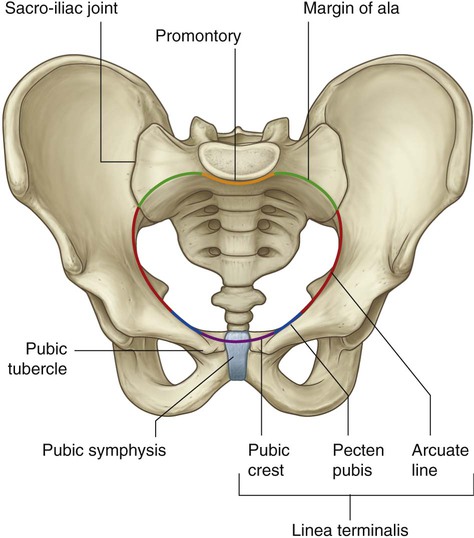
Pelvic Anatomy Posterior / Iliac Screw For Reconstructing Posterior Pelvic Ring In Tile Type C1 Pelvic Fractures Sciencedirect / Über 7 millionen englischsprachige bücher.. Major or minor angles mean pelvic retroversion or anteversion and posterior or anterior pelvic tilt. This quiz is unlabeled so it will test your knowledge on how to identify these structural locations (iliac crest, ischial spine, acetabulum, superior ramus of pubis, posterior superior/inferior iliac spine, lessier. Is divided into posterior (short and long) and anterior ligaments. The pelvic skeleton is formed posteriorly (in the area of the back), by the sacrum and the coccyx and laterally and anteriorly (forward and to the sides), by a pair of hip bones. Major components of the bony pelvis, frontal superior view.
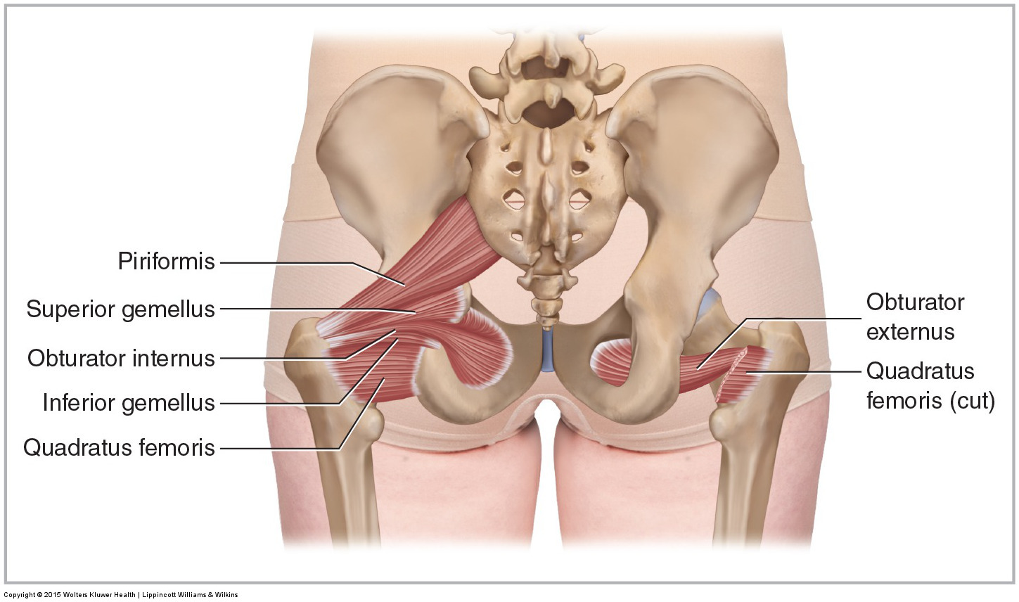
Posterior view of the lumbar spine and pelvis. The anterior division of the internal iliac artery is the main blood supply to the vital organs of the pelvis, namely the bladder (superior vesical artery) and uterus (uterine artery) (figure s5). It can be divided into the greater pelvis and the lesser pelvis. It courses inferiorly, anterior to the piriformis muscle and sacral plexus, extends laterally, and exits the bony pelvis via the greater sciatic notch. During childhood, these sections are separate bones, joined by the triradiate cartilage.
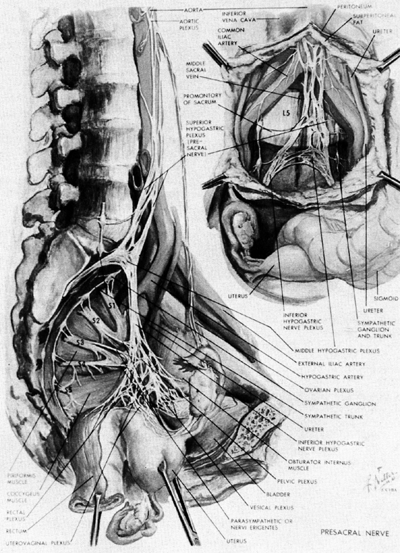
Anatomy Of The Pelvis Glowm from resources.ama.uk.com The left and right sides of the pelvis are joined together by the symphysis pubis (sp) made of cartilage in the anterior, and posteriorly with the sacrum at the sacroiliac (si) joint. In this cadaver study, pelvic organs were resected en bloc, immersed in formalin solution, and transected in the mid sagittal plane. The pelvic bones are smaller and narrower. Injury in pelvic fractures can account for majority of blood loss. The two chains unite in front of the coccyx to form a small ganglion impairment. The inferior hypogastric plexus is also known as the pelvic ganglion. Motions of the joints of the pelvis. The posterior division is of less significance as it pierces the presacral fascia and supplies the gluteal region.
Bones and ligaments of the female pelvis.
(2017, elsevier) should be consulted. The two chains unite in front of the coccyx to form a small ganglion impairment. The inferior hypogastric plexus is also known as the pelvic ganglion. The posterior division is of less significance as it pierces the presacral fascia and supplies the gluteal region. The posterior pelvis normally refers to the bones that make up the rear aspect of the pelvis. Classic anatomical studies have provided few details of the inferior hypogastric plexus morphology or the location and nature of the associated nerves. Major components of the bony pelvis, frontal superior view. At times, it also may refer to structures or tissues found within or attaching to these bones. The orientation of the pelvic inlet the pelvic inlet has an inclination of about 55 to 60 degrees with respect to the anatomical horizontal plane. Pelvis (hip) anatomy quiz for anatomy and physiology! Muscular pelvic floor closure helps to relieve fascial stress. A more detailed account of pelvic anatomy is best found in anatomy texts 1. In this cadaver study, pelvic organs were resected en bloc, immersed in formalin solution, and transected in the mid sagittal plane.
To examine the gross and histologic anatomy of the proximal, mid, and distal posterior vaginal compartment and discuss implications for surgical repair. The pelvic part of the sympathetic chain runs downwards and slightly medially over the body of sacrum, and then along the medial margins of the anterior sacral foramina. The pelvis's frame is made up of the bones of the pelvis, which connect the axial skeleton to the femurs, and therefore acts in weight bearing of the upper body. The paired left and right sacroiliac joints and the symphysis pubis joint. Posterior ligaments provide most of the stability.
Muscles Of The Pelvis from learnmuscles.com The posterior abdominal wall is a complex region of anatomy. The pelvis is a ring structure… The posterior division is of less significance as it pierces the presacral fascia and supplies the gluteal region. In rare cases, it gives rise to a persistent sciatic artery (see later). Runs from the posterolateral aspect of the sacrum and the dorsal aspect of the posterior iliac spine to the ischial tuberosity. The two chains unite in front of the coccyx to form a small ganglion impairment. Major or minor angles mean pelvic retroversion or anteversion and posterior or anterior pelvic tilt. A more detailed account of pelvic anatomy is best found in anatomy texts 1.
Injury in pelvic fractures can account for majority of blood loss.
It courses inferiorly, anterior to the piriformis muscle and sacral plexus, extends laterally, and exits the bony pelvis via the greater sciatic notch. The pelvis consists of the sacrum, the coccyx, the ischium, the ilium, and the pubis. A more detailed account of pelvic anatomy is best found in anatomy texts 1. Major or minor angles mean pelvic retroversion or anteversion and posterior or anterior pelvic tilt. Connects iliac and obturator systems. Is divided into posterior (short and long) and anterior ligaments. Runs from the posterolateral aspect of the sacrum and the dorsal aspect of the posterior iliac spine to the ischial tuberosity. The left and right sides of the pelvis are joined together by the symphysis pubis (sp) made of cartilage in the anterior, and posteriorly with the sacrum at the sacroiliac (si) joint. The inferior hypogastric plexus is also known as the pelvic ganglion. Bones found here include the paired ilium bones of the pelvis and the sacrum and coccyx bones at the base of the spine. In this cadaver study, pelvic organs were resected en bloc, immersed in formalin solution, and transected in the mid sagittal plane. To examine the gross and histologic anatomy of the proximal, mid, and distal posterior vaginal compartment and discuss implications for surgical repair. Major vessels, nerves and organs are located on the inner surface of the posterior abdominal wall.
In rare cases, it gives rise to a persistent sciatic artery (see later). The pelvic ring protects the organs in the lower abdomen from injury. Muscular pelvic floor closure helps to relieve fascial stress. The pelvic skeleton is formed posteriorly (in the area of the back), by the sacrum and the coccyx and laterally and anteriorly (forward and to the sides), by a pair of hip bones. The pelvis is the lower portion of the trunk, located between the abdomen and the lower limbs.
Pelvis And Perineum Clinical Gate from clinicalgate.com The fusion of the pelvic splanchnic nerves, sacral splanchnic nerves, and superior hypogastric plexus along with visceral afferent fibers forms the inferior hypogastric plexus. The posterior pelvis normally refers to the bones that make up the rear aspect of the pelvis. (gilroy et al.) atlas of anatomy 2nd ed., fig. Major or minor angles mean pelvic retroversion or anteversion and posterior or anterior pelvic tilt. This image shows the posterior back view of the female pelvic brim (the bones and ligaments that forms the pelvic region in the female) showing: The pelvic skeleton is formed posteriorly (in the area of the back), by the sacrum and the coccyx and laterally and anteriorly (forward and to the sides), by a pair of hip bones. At times, it also may refer to structures or tissues found within or attaching to these bones. We hope this picture pelvic region posterior view can help you study and research.
The orientation of the pelvic inlet the pelvic inlet has an inclination of about 55 to 60 degrees with respect to the anatomical horizontal plane.
Divides distal and posterior near the si joint into. For more anatomy content please follow us and visit our website: Leads to superior guteal artery and other branches. The pelvis is the lower part of the torso. It is enervated by the obturator nerve. The vertebral column of the lower back includes the five lumbar vertebrae, the sacrum, and the coccyx. Muscles of the pelvic diaphragm, oblique view. The pelvic region is the area between the trunk — or main body — and the lower extremities, or legs. To examine the gross and histologic anatomy of the proximal, mid, and distal posterior vaginal compartment and discuss implications for surgical repair. The fusion of the pelvic splanchnic nerves, sacral splanchnic nerves, and superior hypogastric plexus along with visceral afferent fibers forms the inferior hypogastric plexus. We hope this picture pelvic region posterior view can help you study and research. Each hip bone consists of 3 sections, ilium, ischium, and pubis. Posterior wall of true pelvis medial attachment:
The posterior abdominal wall is a complex region of anatomy pelvic anatomy. The pelvic ring protects the organs in the lower abdomen from injury.